Diagnostic and clinical informatics
Integrated diagnostic solutions for better patient care

Article
The top five things to look for in an enterprise informatics partner
Developing an effective enterprise imaging strategy is often easier said than done. How do you choose a partner who can help you accomplish this? Here are five points to consider when choosing an enterprise informatics partner to focus on providing access to any type of medical image, anywhere, at any time by anyone across the continuum of care in healthcare systems.
Featured solutions in Diagnostic and clinical informatics
- New
Diagnostic Workspace
With our integrated workspace, radiologists can access needed tools, including reporting, advanced visualization analysis and Al-enabled insights. Our solution is designed intuitively to optimize care pathways from orchestration to diagnosis to collaboration, using both DICOM and non-DICOM data with powerful tooling.
NOCTN424PACS -
Cardiovascular Workspace
Cardiovascular Workspace is a scalable and interoperable solution that combines deep clinical expertise with technological innovation to help streamline workflow and improve operational performance throughout the complete cardiovascular care continuum.
NOCTN198 -
PerformanceBridge 4.0
PerformanceBridge* offers an integrated, scalable portfolio of services, tools and support to empower hospital departments to boost their performance and build a program for continuous improvement. It is designed to enable you to focus on enhancing operational efficiency and reducing costs, while maintaining an emphasis on quality, performance and value. PerformanceBridge is a web-based, real-time data analytics platform that aggregates data from multiple sources, such as your HIS, RIS, PACS and financial systems.
896001 -
Radiology Operations Command Center
A multi-vendor, multi-modality, multi-site, backward compatible, safe and secure virtualized imaging support solution that smoothly connects imaging experts in a command center/workspace with technologists at scan locations across their organization.
808001 -
Lung Cancer Orchestrator
The Philips Lung Cancer Orchestrator is an integrated lung cancer patient management system for both CT lung screening programs and incidental pulmonary findings programs that monitors patients through various steps of their lung cancer screening and treatment decision journey. Enhancing confidence with automated tools, the Lung Cancer Orchestrator helps identify and keep more patients – and streamline workflows. Providing a complete solution, the system also comes equipped with the Multidisciplinary Team Orchestrator to help facilitate collaborative clinical decision making.
841017 -
DynaCAD Lung
DynaCAD Lung is a vendor neutral Computer-Aided Detection (CAD) system that provides a robust set of automated tools for radiologists to analyze multi-slice CT exams of the chest. With flexible report templates and automated image registration, DynaCAD Lung helps facilitate synchronous display and navigation of multiple patient exams for initial review and easy follow-up comparison of current and prior study findings.
784022


- 0
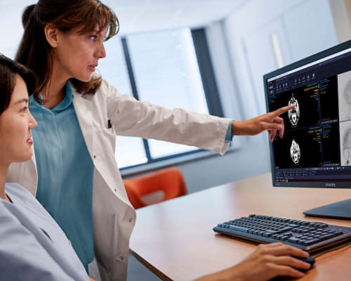
Have access to the complete patient record whenever and wherever needed
A complete radiology workflow with embedded reporting and advanced applications for diagnostic outcomes to eliminate time-consuming manual data searches. A single source of imaging information used to create the Imaging Health Record.
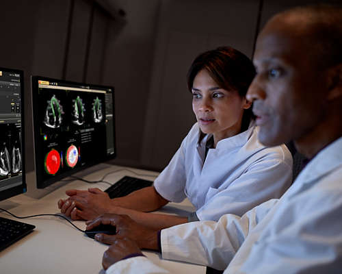
Turn clinical findings into a decisive actionable plan
You need a complete overview of your patient’s cardiovascular history. By seeing the past as well as the now, you can make faster, more informed decisions. Make the leap to integrated cardiology solutions that take sharing data and clinical decision-making to another level.

Revealing timely, actionable insights for precision cancer care
Our oncology informatics solutions enable generation, interpretation, and integration of the right insights at the right time across specialties, delivering insights directly to oncology care teams.
Unify data, unlock actionable insights
Empower your staff to improve operational outcomes and reduce costs, in real time. Philips PerformanceBridge provides access to aggregate data and AI generated best-case scenarios, predictive budgeting, staffing mix/modelling, and right-size fleet projections.
Key capabilities
-
![Integrated Diagnostics]()
Unleash the power of fully integrated solutions. Discover our user-friendly, clinically proven portfolio of integrated diagnostic informatics solutions.
-
![Echocardiography]()
Echocardiography solutions from Philips Healthcare.
-
![Philips Healthcare for clarity on the cancer care journey]()
Philips helps bring more clarity at every moment of cancer care through streamlined multidisciplinary workflows and integrated patient data. See how.
-
![Cardiac imaging]()
Our cardiac imaging solutions provide excellent image quality and streamlined analysis, reporting and insights for a more confident diagnosis.
-
![Philips radiology workflow solutions]()
Learn about our portfolio of integrated radiology imaging workflow solutions and how they can reduce exam times, minimize the need to rescans and lead to fast reporting of critical findings.
Footnotes
Product may not be available in all geographies. Please check with your local Philips representative to ascertain applicability of this solution for your region and language requirements.